At Greenspoint Dental in Houston, Texas, we are proud to provide exceptional, gentle general dentistry alongside our leading-edge cosmetic, restorative, and orthodontic dental services.
If you, or someone you care about, needs comprehensive, compassionate dental care, you can visit our clinic.
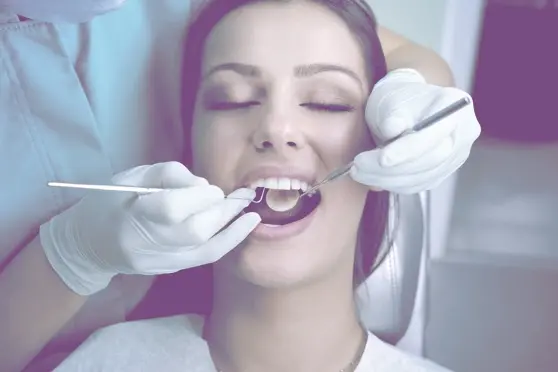
Many of our Houston area patients have come to us after a previous experience left them anxious about their dental needs. Once they experience our gentle touch dentistry, they leave feeling more comfortable about their future dental care and visits.
We pay attention to all your needs by offering pleasing amenities such as anxiety-reducing music, comfortable lighting and chairs, soft warm blankets, pillows, and more.
Whether you need nitrous oxide (also called laughing gas), Novocaine, or even full sedation dentistry so you can sleep through the entire procedure, we are here to help. Our dedicated team will work to provide superior dental care to ensure that you feel comfortable, confident, and satisfied at every step.
Come See Houston’s Best Dental Team
Our general dentistry in Houston includes a wide range of procedures and services which promote oral health and help to preserve your natural teeth. Good oral health starts with healthy habits in childhood, including regular exams, dental sealants, orthodontic evaluation around age 8, and other preventative procedures, and continues over a lifetime.
As you age, your oral needs can change. From fillings to oral cancer screenings, we are here at every life stage to improve and protect your very best smile.
Our General Dentistry Services in Houston
Bi-annual Checkups and Cleaning
Bi-annual checkups and cleanings are an important part of your oral healthcare. Regular exams can catch small issues before they become bigger ones. Having a six-month cleaning removes excess tartar. This is important because it can help prevent gum disease.
Tartar, also called calculus, is actually a form of hardened dental plaque caused by a build-up and calcification of minerals. When tartar accumulates on the teeth, it dramatically increases your risk of developing gingivitis (inflammation of the gums). Because tartar bonds and hardens (calcifies) on teeth, it must be removed by a trained dental professional.
Dental crowns are like a cap that is custom made to fit onto the top of your tooth. A crown can restore and strengthen the integrity of a damaged tooth. Sometimes a crown is required when a cavity is too large for a filling. In other cases, a tooth may be chipped, weakened or worn down, or badly shaped.
A crown may also be used to improve the appearance or save a tooth after a root canal. A properly made dental crown matches the shade of your teeth, appears and functions like a natural tooth, and fits comfortably in your mouth, and works with your bite. Generally speaking, a crown usually takes one to two visits to complete.
Dental fillings have come a long way in recent years. New composite materials are not only stronger, they last longer and can be color-matched to your teeth to make them less noticeable.
Inlays and Onlays are sometimes called indirect filling because they are crafted before being fitted and bonded onto the tooth. An inlay covers the chewing surface of the tooth, including the nooks and fissures of the tooth. An onlay, on the other hand, covers the cusps, or mounded sections of the tooth.
Dental sealants create a barrier that protects your teeth from dental plaque and acids caused by the bacteria in your mouth. Dental sealants are painted onto the surface of the teeth to reduce the chance of dental decay. They are often used to protect the teeth of children, although they can be used at any age. Regular dental care is still required, but properly applied dental sealants can go a long way in protecting your teeth.
Oral Cancer Screening
Although oral cancer is rare, the odds of developing oral cancer do increase with age. Spotting oral cancers early improves the odds that treatment will be successful. Studies have shown that dental professionals are often the first to spot oral cancer in patients.
Gentle General Dentistry for Those in the Houston Area
At Greenspoint Dental, we are known for gentle, yet sophisticated dental care. You will see the difference from the moment you step into our spacious, well-appointed office. You’ll notice when our caring dentists put you at ease while you are in the chair. From start to finish, we are dedicated to your comfort and care.
If you live or work in the Houston area and would like to schedule a free consultation at our general dentistry, please call (281) 823-9987 or click here to schedule an appointment today.